RVG (rheovasography) of the lower extremities: features, principle of carrying out, decoding and norms
Many severe pathologies originate in,seemingly small, minor manifestations, to which very few people pay attention. Even mild vascular changes can subsequently lead to a stroke, a heart attack, amputation of the extremities. Vascular diseases occur for various reasons, these can be psychoemotional stresses, unbalanced diet, intense rhythm of life. The earlier an unstable condition is detected, the onset of the disease, the more literate and adequate the therapy is selected, and this is an important aspect for maintaining health. In medicine, preference is given to noninvasive methods of research, which are of great informativeness, one of them is reovasography of the lower extremities (RVG). We will become acquainted in more detail with this method: for what purpose is carried out rheovasography, to whom is shown and how the results of the survey are deciphered.

The concept of rheovasography
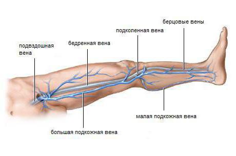
Reovasography is one of the waysdiagnosis, in which the circulation of blood in the lower and upper limbs of the patient is examined. The blood circulation, studied in this way, is called hemodynamics. She reveals the picture of the condition at the time of examination of the cardiovascular system, and also allows to assess the tone of the vessels. RVG shows the condition of the arteries, veins in the selected area of the legs, hands, makes it possible to determine the occurring changes in the walls of the vessels. When deciphering it is seen if there is a partial narrowing of the vessels or there is complete obstruction.
In the life of a person the main loadaccount for the lower extremities, so most often the study is assigned to this part, this allows you to evaluate the blood flow of the vessels of the legs and to prescribe the right treatment.
It is possible to go through re-printing not only bydirection of the doctor, but also independently. This research is carried out in many specialized clinics at an affordable cost. If you need rheovasography of the lower extremities in Moscow, you can contact the following addresses:
- 2-nd Tverskoy-Yamskaya Pereulok, Building 10 - OJSC "Medicine".
- Banniy lane, house 2 - SvsR.
- Mkrn Novogorsk in Khimki - Design Bureau No. 119.
- Ul. Novozavodskaya, house 14a - polyclinic № 2.
- Ul. Lyapidevskogo, d. 14/1 - "Dobromed".
- Ul. Budayskaya, house 2 - JSC "Russian Railways", Central Design Bureau No. 2.
Depending on which institutionthe procedure is carried out, its reputation, location, from the quality of equipment on which rheovasography of the lower extremities passes, the price may fluctuate. The cost is from 800 to 2500 rubles.
Reovasography is one of the non-invasive methodsexamination where no penetration into the body is required. For carrying out use a high-frequency current. This study is not used for the vessels of the head, in this case the diagnosis is called rheoencephalography.

Procedure
For carrying out rheovasography of vessels of the upper andlower extremities, an electrocardiograph (ECG apparatus), special electrodes and a rheographic attachment are needed. The task of a medical professional is greatly facilitated if computer registration of indicators is used. The study uses a high frequency current that does not bring any unpleasant sensations. Blood can conduct an electric current, for this reason, the resistance varies in different phases of the blood flow. The dependence here is inversely proportional: with more blood filling - less resistance of tissues. From high indicators to low there is a constant fluctuation. The reovasogram graphically displays the passage of pulse oxen, the amplitude increases with increasing blood filling and, conversely, decreases when it decreases. The doctor of functional diagnostics sees a violation on the chart and can draw conclusions about the nature of the blood flow.
Indications for examination

Reovasography of the vessels of the lower extremities is used both for the initial study of vascular pathologies and for monitoring the development of the disease. The indications are:
- Atherosclerosis, which often contributes to the disruption of proper nutrition of tissues and the narrowing of lumens in the arteries.
- Diabetes. Rheovasography of the upper and lower extremities is performed. This ailment can lead to micro- and macroangiopathies, which subsequently develop a diabetic foot.
- Raynaud's disease. Typical neurocirculatory asthenia, periodic spasms of peripheral arteries are possible.
- Expressed headaches that arise due to violations of the outflow of venous blood, as well as spasms of arterial vessels.
- Chronic brain pathologies, intracranial hypertension.
- Neurocirculatory asthenia, which depends on the lability of vascular tone.
- Diseases of the venous channel, which contribute to violations of outflow of blood, in particular through the veins of the legs.
- Thrombophlebitis, leading to the formation of blood clots on the walls of blood vessels.
- Phlebeurysm.
- Embolism, when there is a blockage in the bloodstream.
- Obliterating endarteritis, which can lead to lameness.
In the absence of pronounced diseases, RVG is recommended in cases of numbness of the limbs, with the appearance of blue eyes and seizures, with loss of sensitivity, skin discoloration, swelling.
Pros of the survey
Each study has its own advantages,and shortcomings. They can be connected with the technical side or with the professionalism of the staff. It is important that all the negative sides can be adjusted and debugged the diagnostic process for the better. Pros of the RVZ are as follows:
- Non-invasive method. A significant positive point - the patient is not subjected to unpleasant and painful effects, for example, catheterization of veins. For the study does not require a mode specially equipped cabinet, enough necessary devices.
- Ease of carrying out. A big plus for the staff. The rheovasogram can be removed by a qualified nurse, the task of it consists in applying electrodes and including a resograph. The results will be analyzed by the doctor.
- Inexpensive and affordable research equipment.
- Safety procedures. RVG is possible for children and pregnant women.
- Reliable information. With great accuracy, the patency of the vessels, the quality of the outflow of blood, pathology and the level of their prevalence are determined.
- It is possible to carry out a differential diagnosis of vascular damage from a functional disorder.

Minuses
Reovasography of the lower extremities has very small disadvantages, the following points can be assigned to them:
- Human factor. If there are too many patients, the lack of proper rest at the nurse can lead to the dissipation of care, and this can affect the quality of registration. If the working conditions, the rest regime are respected, the qualification of the medical staff is high, then this minus is absent.
- Quality of technology. Rheographic attachments can interfere, which affects the results. The use of computers excludes such shortcomings in the study.
Preparing for rheovasography
Preparation for reovasography of the lower extremities is not at all complicated, but it is necessary to comply with all the requirements that are required before the study.
- 15 minutes before the test, the patient should completely relax, get a preliminary rest.
- Those who smoke, in two hours should completely exclude the flow of nicotine into the blood.
- If there is a course of treatment with any medications, one day before the RRT it is necessary to abandon the drugs.
- When passing the rheovasography, the limbs should be freed from clothing.

Conducting research
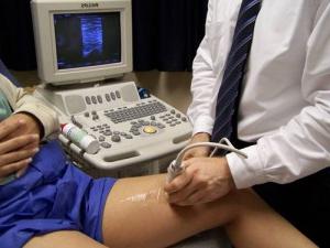
The patient lies on the couch, the pose on the back. The skin of the extremities is degreased with alcohol. Sensors are attached to the treated areas. From the device wires stretch, end with electrodes, which will read the information. Signals are transmitted to the screen, the rheovasogram is recorded, the main indicators are calculated.
The rheovasogram can be performed usingmultichannel rheographs, or the investigation is carried out sequentially, beginning from the sites remote from the center ones, ending with the neighbors. A characteristic feature when placing conductors is strict symmetry.
Indicators
Rheovasography of the lower extremities is performed withthe purpose of studying the quantitative indicators of the state of blood vessels. Synchronous waves of readings are equated to the pulse rate. By waves, one can judge the vascular filling, depending on the phase of cardiac contraction in a certain time period. As a result of the study, the RI (rheographic index) is output. It is calculated by comparing the amplitude of the waves with the height (the calibration pulse). The value of RI directly depends on the filling of blood vessels with blood, with its help it is possible to calculate the total intensity with which the organ is filled with arterial blood. The following parameters also include the main parameters: the outflow value; elasticity; peripheral resistance.

Rheovasography of vessels of the lower extremities (decoding)
After receiving the rheovasogram, time arrivesdeciphering, which is carried out by a qualified specialist. This is the main point of the study, all pathologies are found here and the question of further treatment is being decided. The interpretation of rheovasography of the lower extremities includes the following important points:
- The main focus is the study of RI. A value of 0.04 indicates a sharp decrease in the norm, an index of 0.04-0.05 - a moderate decrease. Normal RI is considered to be above 0.05.
- The index of elasticity (IE). The sharply lowered figure is less than 0.2. Moderately reduced - 0,2-0,4. Normal - 0,4.
- To determine the outflow of blood in the vessels, calculate the corresponding index, the norm of which is 0.2-0.5. If the value is less, the outflow is facilitated; the indicator above 0.5 indicates difficulties in outflow.
- Parameters of peripheral resistance: more than 0.55 - overestimated; less than 0.15 - understated. The norm is 0.2-0.45.
Functional tests

To identify hidden pathologies, functional pharmacological tests can be used:
- Nitroglycerin. After resorption of nitroglycerin, RVG is performed, after which the result is compared with the usual one. This is necessary in order to distinguish between organic narrowing and functional spasms.
- Compression. Used in the study of deep vein thrombosis. For comparison with the usual indices of RVG, the cuff on the hip is superimposed and the indicators are fixed after removal.
- Cold. It is carried out for the diagnosis of various pathologies in the hands. Cold water is used. The rheogram is taken three times after a certain time, the results of the restoration of the blood flow are compared, conclusions are drawn. Negative test - to restore RI in the 7th minute. A positive test is a slow recovery of up to 30 minutes.


