The mechanism of inhalation and exhalation. Structure and general patterns of the functioning of the respiratory system
Without breathing, a person will not survive for seven minutes. This is the most important function of the body, although we do not make much effort to implement it. How does the inspiration and expiration mechanism work? What organs and systems does it use?
Breathing Rights
For life, we need oxygen. This is the key element of respiration, which ensures the metabolism and energy in the body. It enters the lungs in the gaseous state together with air, spreading throughout the body, where it is oxidized and excreted from the body in the same way.
The breathing mechanism - inhalation-exhalation - workscontinuously. At the moment a person makes about 14 such movements, in infants the number increases to 50. The breath of a person is one of the few processes that can be managed consciously and unconsciously.
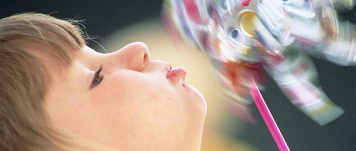
By a small effort of will we can regulate itfrequency and duration, and if necessary, and completely delay it for a few seconds. This skill is necessary for a person, for example, during a voyage. Too long to hold our breath we are not capable, the brain without oxygen dies down within five to seven minutes.
Mechanism of inspiration and expiration
Living beings have different ways andmechanisms of respiration. Some use the entire surface of the body, others use gills, others have lungs. A person has internal tissue and external pulmonary respiration. Tissue represents oxygen consumption by cells of internal organs.
Pulmonary respiration is carried out in two stages: gas exchange with the alveoli, and then with blood. Air from the atmosphere, saturated with oxygen, passes through the nasopharynx, larynx, tracheobronchial tree and enters the pulmonary alveoli.
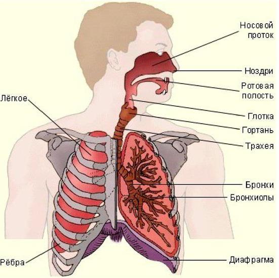
They give atmospheric air to the blood, whichcarries it to all organs along the vessels. From the blood into the alveoli gets air, saturated with carbon dioxide, which is excreted from the body together with the exhalation.
The inhalation and expiration mechanism provides ventilationalveoli. It is carried out with the help of the respiratory musculature, which expands the thorax, allowing to take in the lungs up to 7 liters of air per minute. The increase occurs by raising the ribs (more often in women) or by flattening the diaphragm (in men, as well as under physical exertion).
Upper respiratory tract
The importance of respiratory organs is not the same, eachof them have their own functions. The human respiratory system includes the upper and lower respiratory tract, and the respiratory system proper. The upper paths are represented by the nasal cavity, nasopharynx, oropharynx and partly the oral cavity.
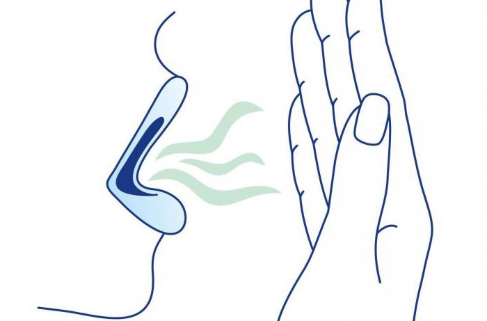
The inside of the nasal cavity is coveredmucous membrane and hairs. It acts as a filter, the main task of which is to prevent dust, dirt and microbes from entering the body. Here the air is warmed and moistened.
Two channels of the nostrils connect the cavity with the nasopharynx. It, in turn, is connected to the Eustachian tube, responsible for equalizing the pressure.
In the oropharynx, respiratory and foodways. It is limited to the posterior and lateral walls of the oral cavity and is responsible for clear pronunciation. During eating and speaking, the soft sky rises, preventing food and air from entering the nasopharynx.
Lower respiratory tract
The pharynx carries the air to the larynx. It starts from her lower respiratory tract. The larynx is formed by paired and unpaired cartilages, connected together by ligaments and muscles. The contraction of muscles changes the shape of the glottis and the tension of the ligaments, resulting in the formation of sounds.
The larynx is connected to the tube up to 15 centimeters inlength - trachea. Its end splits, passing into the bronchi. The main function of the trachea is the passage of air into the lungs and back. It is mobile and consists of cartilage, so the air passes through it at any turns of the neck.
Bronchi are a paired organ and enter the lungs. The left bronchus is thinner than the right one, the right bronchus is more vertical. They are formed by cartilaginous rings and smooth muscles, inside are covered with a mucous membrane.
Each of them has ramifications - in their righteleven, in the left ten. Lymph nodes in the fork transport lymph from the lung tissue, the blood is transferred to the bronchial arteries from the thoracic aorta.
Lungs
The lungs are sometimes referred to as the lower respiratory tract. They are in the chest cavity from the left and right sides of the heart, and their base is located on the diaphragm. Outside, the lungs are covered with the pleura and the pleural sac. Between them is a lubricating fluid that prevents friction.
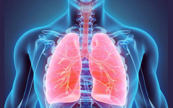
Lungs consist of several segments (right ofthree, the left of the two), which are divided into ten smaller shares. Inside of them are located bronchi, which, in turn, are divided into small bronchioles, acini, and end with alveolar sacs.
Bags represent numerous alveoli - globular formations, braided by capillaries. In an adult, their number is about 700 million. They are responsible for gas exchange.
From them blood enters the blood vessels,oxygenated. Blood along the arteries moves directly to the heart, and along the way it spreads across all tissues and organs. In return, they give blood saturated with carbon dioxide, which returns through the veins to the alveoli to exit through the lungs, bronchi, trachea, pharynx back into the atmosphere.
Exercising breathing
The inspiration and exhalation mechanism is controlled by the centerbetween the posterior and oblong brain. Receptors that regulate the process of breathing are located on the walls of the bronchi. The movement of air is also due to the difference in pressure: when it is inhaled, it is below atmospheric pressure, and when exhaled, it is vice versa.
Lungs can skip up to 5 thousand millilitersair during inspiration and exhalation. But with normal breathing, the volume is only 500 milliliters. The maximum inhalation can be about 2500 ml.
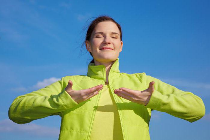
Man does not breathe absolutely all the air. Part of it lingers in the alveoli, so that the ratio of oxygen and carbon dioxide adheres to the same level. This is the functional residual capacity of the lungs.
In the process of breathing, various groupsmuscles, depending on the person's occupation. The diaphragm participates during sports training or physical exertion, when the abdominal area strains. In a calm state a large role is assigned to the intercostal muscles.
</ p>


